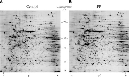
FIGURE 1.

Silver-stained 2-dimensional gel images representing total proteins extracted from human duodenal mucosal biopsy samples after maltodextrin (A) and protein-supplemented maltodextrin (B) enteral perfusion. Differentially expressed proteins (i.e., at least ±1.5-fold modulated; circled spots with a number) were determined by statistical analysis (paired Student’s t test, P < 0.05) and correspond to the samples analyzed by liquid chromatography–tandem mass spectrometry. Protein identification results are depicted in Table 1. pI, isoelectric point; PP, protein powder.
