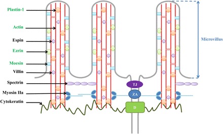
FIGURE 4.

Intervention levels of ezrin, moesin, plastin 1, and β-actin in the apical structure of human duodenal epithelial cells. Proteins in green are upregulated after protein supplementation. These modifications suggest an actin cytoskeleton remodeling in human duodenal mucosa. D, desmosome; TJ, tight junction; ZA, zonula adherens.
