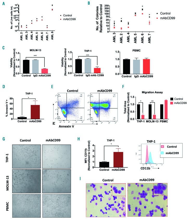
Figure 7.
Effect of CD99 mAB on acute myeloid leukemia (AML) cells. (A) Cell viability of AML blasts treated with 20µg/mL of mAbCD99 for 48 hours (h) and measured using trypan-blue (n=7). (B) Total number of colonies comparison between AML blasts treated with mAb CD99 and control blasts (n=6). (C) Viability of THP-1, MOLM-13 and healthy donor peripheral blood mononuclear cells (PBMC) treated with 5µg/mL of mAbCD99 for 48 h and measured using alamar blue. (D and E) Apoptosis measured in THP-1 and MOLM-13 cells treated with mAbCD99 for 72 h and stained with Annexin V for flow cytometry analysis. (F and G) Quantitative analysis and representative images of migration of THP-1, MOLM-13 and PBMC treated with mAB CD99 towards SDF-1α performed in a transwell plate. (H) CD11b measured by flow cytometry in THP-1 cells 72 h post treatment with 2.5 µg/mL of mAB CD99. (I) Representative images for Wright-Giemsa staining of THP-1 cells treated with 2.5 µg/mL of mAb CD99 for 72 h. (***P<0.001; **P<0.01; *P<0.05).

