
Fig. 1.
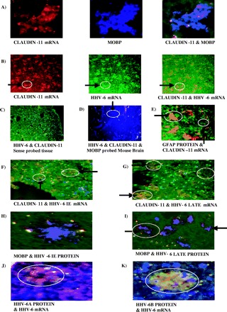
Both immediate early and late viral gene expression is seen in all samples and locates to the cytoplasm of oligodendrocytes, indicating an active infection. The number of cells that are positive for HHV-6 mRNA varies but cells tend to be seen in clusters. (A) Representative FISH of claudin-11 mRNA (rhodamine red), ISH for myelin oligodendrocyte basic protein (MOBP) using AMCA (blue)-conjugated secondary antibody with the two colour images merged to demonstrate co-localization of both signals indicating that the mRNA probe correctly identifies oligodendrocytes (magnification ×400). The image is taken from normal control tissue. (B) Representative double FISH for claudin-11 mRNA (rhodamine red), HHV-6 mRNA (fluorescein) and the two colour merged image demonstrating co-localization of both signals to oligodendrocyte cells (orange). The circle and arrow indicate two representative cells showing first claudin-11 mRNA, then HHV-6 mRNA and finally the two merged images (magnification ×200). The image is taken from multiple sclerosis NAWM tissue. (C) Normal control tissue probed with sense probes for HHV-6 (fluorescein) and claudin-11 (rhodamine red). All that can be seen are the DAPI-counterstained nuclei (magnification ×200). (D) Mouse brain section probed for HHV-6 mRNA (fluorescein), claudin-11 mRNA (rhodamine red) and MOBP protein (AMCA secondary). Only MOBP signal is present. The circle and arrow indicate a representative oligodendrocyte (magnification ×200). (E) Representative ISH for GFAP (fluorescein secondary) and claudin-11 mRNA FISH (rhodamine red). The signals do not co-localize, the circle and arrows highlighting examples of both (magnification ×200). The image is taken from normal control tissue. (F) Representative double FISH for claudin-11 and HHV-6 immediate early mRNA with both signals co-localizing (orange). The circle and arrow indicate two representative cells where the two mRNA signals co-localize (magnification ×200). (G) Representative double FISH for claudin-11 and HHV-6 late mRNA with both signals co-localizing (orange). The circle and arrow indicate two representative cells where the two mRNA signals co-localize (magnification ×200). (H) Representative double ISH for MOBP (AMCA secondary) and HHV-6 immediate early (rhodamine secondary) proteins exhibiting co-localization of the signal to oligodendrocytes (purple) confirming mRNA FISH results (magnification ×400). (I) Representative double ISH for MOBP (AMCA secondary) and HHV-6 late (rhodamine secondary) proteins exhibiting co-localization of signal to oligodendrocytes (purple) confirming mRNA FISH results. The circle and arrow indicate two representative cells where the two protein signals co-localize (magnification ×200). Images F–I are from from multiple sclerosis lesion tissue. (J) Representative ISH for HHV-6A protein (rhodamine secondary) and HHV-6 mRNA FISH (fluorescein) (magnification ×400). The image is taken from multiple sclerosis NAWM. (K) Representative ISH for HHV-6B protein (rhodamine secondary) and HHV-6 mRNA FISH (fluorescein) (magnification ×400). The image is taken from multiple sclerosis NAWM.