
Figure 1.
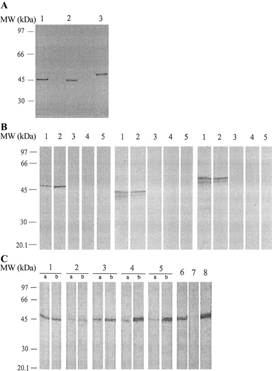
A, Western blot analysis of the expressed recombinant nucleocapsid proteins with anti-His monoclonal antibody. Each protein had a 6 histidine tag at the N-terminal end. Western blot results showed clear and specific bands at molecular weight (MW) positions 47, 44, and 50 kDa, corresponding to the recombinant nucleocapsid proteins of severe acute respiratory syndrome (SARS)—Cassociated coronavirus (CoV) (lane 1), human CoV (HCoV)—229E (lane 2), and HCoV-OC43 (lane 3). B, Western blot analysis of the antigenicities of recombinant nucleocapsid proteins. Left, Nucleocapsid protein of SARS-CoV with SARS-CoV—immune rabbit serum (lane 1), monoclonal antibody to the nucleocapsid protein of SARS-CoV (lane 2), HCoV-229E—immune rabbit serum (lane 3), HCoV-OC43—immune rabbit serum (lane 4), and nonimmune rabbit serum (lane 5). Center, Nucleocapsid protein of HCoV-229E with HCoV-229E—immune rabbit serum (lane 1), monoclonal antibody to the nucleocapsid protein of HCoV-229E (lane 2), SARS-CoV—immune rabbit serum (lane 3), HCoV-OC43—immune rabbit serum (lane 4), and nonimmune rabbit serum (lane 5). Right, Nucleocapsid protein of HCoV-OC43 with HCoV-OC43—immune rabbit serum (lane 1), monoclonal antibody to the nucleocapsid protein of HCoV-OC43 (lane 2), SARS-CoV—immune rabbit serum (lane 3), HCoV-229E—immune rabbit serum (lane 4), and nonimmune rabbit serum (lane 5). All bands appear at the proper MW position. C, Western blot analysis of the HCoV-229E culture filtrates with paired serum samples from patients with SARS (lanes 1—5), HCoV-229E—immune rabbit serum (lane 6), HCoV-OC43—immune rabbit serum (lane 7), and monoclonal antibody to the nucleocapsid protein of HCoV-229E (lane 8). Lanes 4b (patient 4) and 5b (patient 7), which show results of convalescent-phase serum samples from 2 patients with SARS who had high titers in the HCoV-229E nucleocapsid protein—based ELISA, exhibit prominent immunoreactive bands at ∼44 kDa. Reactions with specific monoclonal antibody to the nucleocapsid protein of HCoV-229E are visible at the same MW. Three pairs of acute- and convalescent-phase serum samples had the same reactive band at ∼44 kDa (lane 1, patient 10; lane 2, patient 8; lane 3, patient 3). All bands appear at the proper MW position.