
Fig. 7.
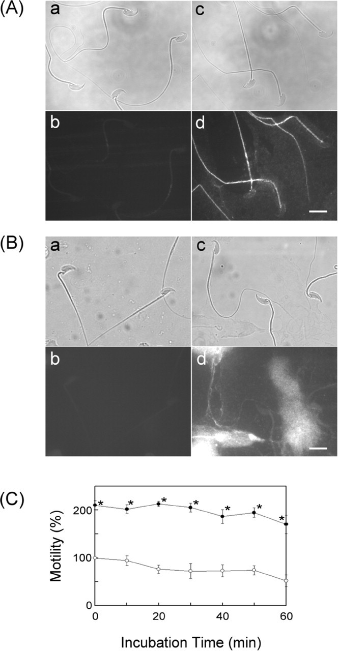
Analysis of sperm motility under the influence of CEACAM10. A) Demonstration of the CEACAM10-binding zone on epididymal spermatozoa. Fresh sperm were incubated with or without CEACAM10 as described in Materials and Methods. The cells on slides were incubated with normal serum (a and b) or affinity-purified anti-CEACAM10 antibody (c and d). The slides were then incubated with rhodamine-conjugated anti-rabbit IgG and observed via light microscopy (a and c) or fluorescence microscopy (b and d). Bar = 10 μm. B) Illustration of CEACAM10 on ejaculated sperm. Freshly prepared cells (see Materials and Methods) on slides were incubated with normal serum (a and b) or the CEACAM10 antibody (c and d) and followed by incubation with rhodamine-conjugated anti-rabbit IgG. The slides were observed via light microscopy (a and c) or fluorescence microscopy (b and d). Bar = 10 μm. C) Freshly prepared mouse spermatozoa in modified Tyrode solution (105 cells/ml) containing 1.8 mM CaCl2 were incubated alone (○) or in the presence of 90 μM CEACAM10 (•) at 37°C for 0 to 60 min. Cell motility determined at each specified incubation time was expressed as a percentage of control cell motility at time zero. Points are mean ± SD for three determinations. *P < 0.01 in a paired statistical comparison with the corresponding control. Values were evaluated using one-way analysis of variance