
Figure 2.
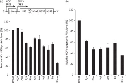
Anti-HCV effect of MOP AADs in HCV subgenomic replicon cells. (a) Schematic diagram of an HCV subgenomic replicon RNA (top panel). The R-1 cells that support constitutive replication of an HCV subgenomic replicon, depicted in (a), were left untreated (DMSO) or treated with the indicated MOP AADs (10 μM) or 100 IU/mL IFN-α for 48 h. Thereafter, whole-cell lysates were prepared and subjected to western blot analysis using an anti-NS5B antibody. Expression of α-tubulin was used to control for equal protein loading. The normalized level of NS5B was determined by densitometric analysis of immunoblots. The relative level of NS5B, expressed as the percentage of the DMSO-treated control, is shown. (b) R-1 cells were treated with 10 μM of the indicated, selected MOP AADs for 48 h. The intracellular HCV genome copy number was quantified by real-time qRT–PCR and expressed as the percentage of the DMSO vehicle-treated control cells.