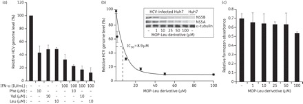
Figure 3.

Anti-HCV activity of the selected MOP derivatives alone or in combination with interferon in HCV (genotype 2a)-infected cells. (a) Huh7 cells grown to ∼60% confluency in 6 cm plates were infected with genotype 2a HCV (JFH1) for 4 h in serum-free medium. Then, cells were washed and complete medium was added. Cells were treated with 10 μM MOP AADs alone or in combination with IFN-α (100 IU/mL) for 72 h. The intracellular HCV genome copy number was quantified by real-time qRT–PCR as in Figure 2(b). (b) Huh7 cells were treated with the indicated concentrations of the MOP-Leu derivative for 72 h before measurement of HCV genome copy number to determine the IC50 value. The inset shows the steady-state expression levels of NS5A and NS5B analysed by western blot of HCV-infected cell lysates treated with MOP-Leu. The NS5A and NS5B levels were determined by densitometric analysis of immunoblots that were normalized to α-tubulin levels. (c) Cytotoxicity of MOP derivatives against Huh7 cells was measured by the MTT assay. Error bars represent standard deviations of triplicates per condition.
