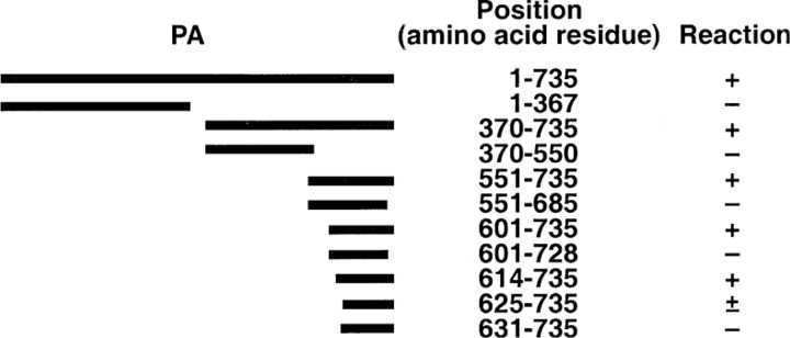
Figure 5.
Summary of epitope mapping of anti–protective antigen (PA) W2 antibody by radioimmunoprecipitation assay. 35S-labeled PA peptides, prepared in vitro, were incubated with anti-PA W2. The immune complexes were captured by protein G–coupled agarose beads and were separated by SDS-PAGE. The PA peptide was detected by exposing the dried gel to an x-ray film. Numbers denote the starting and ending amino acids. The peptides that reacted with antibody and, hence, were detected on an x-ray film were considered to be positive (+). Faint intensity of the band on an x-ray film denoted a partial reaction (±)

