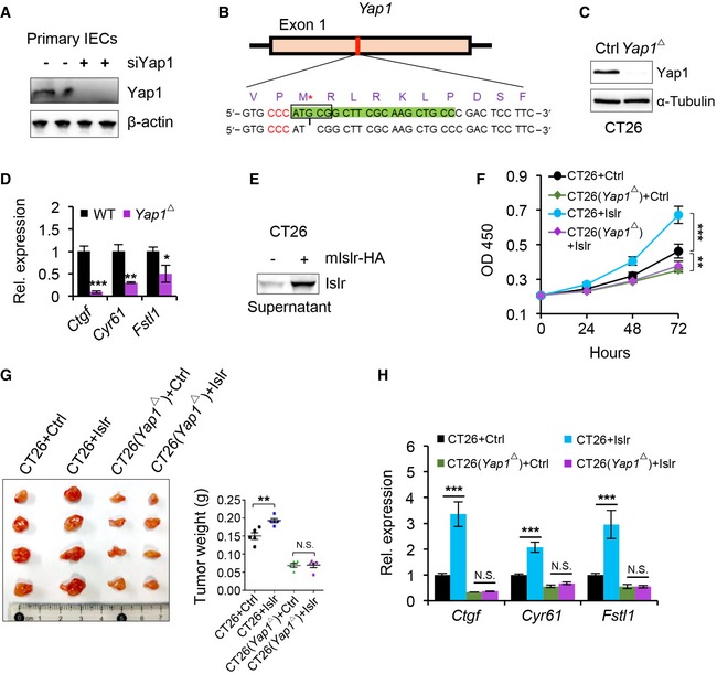
Figure EV5. Deletion of Yap1 abrogated the mIslr‐mediated promoting effect of tumor growth.

- Western blotting for Yap1 in primary intestinal epithelial cells 24 h after Yap1 siRNA treatment. β‐actin was used as a loading control. The sequences of Yap1 siRNA are in Appendix Table S5.
- Generating Yap1‐deficient CT26 CRC cells using Crispr/Cas9 technique. gRNA marked by green color. Protospacer adjacent motif (PAM) sequence marked by red color. G deletion is indicated by an asterisk.
- Western blotting for Yap1 in normal and Yap1‐deficient (Yap1 Δ) CT26 CRC cells. α‐Tubulin was used as a loading control.
- qRT–PCR for Ctgf, Cry61, and Fstl1 in normal and Yap1‐deficient (Yap1 Δ) CT26 CRC cells; n = 3.
- Western blotting for mIslr in the supernatant from CT26 CRC cells transfected with pcDNA3.1 or pcDNA3.1‐mIslr plasmids.
- The growth curve of CT26 CRC cells under indicated conditions; n = 5.
- Gross images of xenografted tumors 10 days after transplantation. Quantification of tumor weight; n = 5.
- qRT–PCR for Ctgf, Cry61, and Fstl1 in CT26 CRC cells at indicated conditions; n = 3.
