-
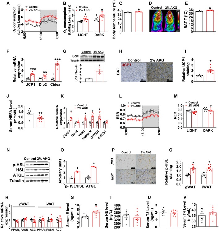
A, B
Oxygen consumption in male C57BL/6 mice after 11 weeks of AKG supplementation (n = 8 per group).
-
C
Body temperature of male mice after 11 weeks of AKG supplementation (n = 9 per group).
-
D, E
Representative images (D) and quantification (E) of BAT thermogenesis induced by 6‐h cold exposure at 4°C in male mice supplemented with AKG for 11 weeks (n = 9 per group).
-
F, G
The mRNA expression of thermogenic genes (F) and immunoblots and quantification (G) of UCP1 protein in BAT of male mice after 11 weeks of AKG supplementation (n = 3–6 per group).
-
H, I
DAB staining (H) and quantification (I) of UCP1 in BAT of male mice supplemented with AKG for 11 weeks (n = 9 per group).
-
J
Serum levels of NEFA in male mice supplemented with AKG for 11 weeks (n = 9 per group).
-
K
The mRNA expression of CD137, CD40, TBX1, TMEM26, CITED1, and slc27a1 in iWAT of male mice supplemented with AKG for 11 weeks (n = 8 per group).
-
L, M
Respiratory exchange ratio (RER) in male C57BL/6 mice after 11 weeks of AKG supplementation (n = 8 per group).
-
N, O
Immunoblots (N) and quantification (O) of p‐HSL and ATGL protein in gWAT of male mice after 11 weeks of AKG supplementation (n = 3 per group).
-
P, Q
Representative images (P) and quantification (Q) of p‐HSL DAB staining in gWAT and iWAT of male mice after 11 weeks of AKG supplementation (n = 9 per group).
-
R
The mRNA expression of PPARγ, FASN, and ACC in the gWAT and iWAT from male mice supplemented with AKG for 11 weeks (n = 6 per group).
-
S–V
Serum levels of E (S), NE (T), T3 (U), and T4 (V) in male mice supplemented with AKG for 11 weeks (n = 8–9 per group).
Data information: Results are presented as mean ± SEM. In (B, C, E–G, I–K, M, O and Q–V), *
‐test.

