
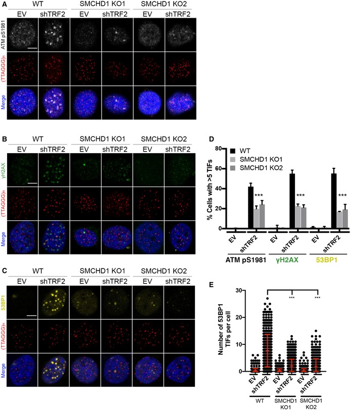
Figure 4. SMCHD1 promotes TIF formation and stimulates ATM signaling from TRF2‐depleted telomeres.
-
A–CRepresentative images for detection of ATM pS9181, γH2AX, and 53BP1 at telomeres in wild‐type (WT) and SMCHD1 knockout HeLa cells transfected with shTRF2 plasmid and empty vector (EV) control. Immunofluorescence (IF) for ATM pS1981 (gray), γH2AX (green), and 53BP1 (yellow) was combined with telomeric (CCCTAA)3‐FISH (red) and DAPI staining total DNA. Scale bars: 5 µm.
-
DQuantification of the number of cells containing > 5 telomere dysfunction‐induced foci (TIFs) detected as in (A‐C). Data represent the mean of four independent experiments ± SD (> 200 cells/condition/experiment) for ATM pS1981 and three independent experiments ± SD (> 200 cells/condition/experiment) for γH2AX and 53BP1. ***P < 0.001; unpaired two‐tailed Student's t‐test.
-
EQuantification of the number of 53BP1 TIFs per cell detected as in (C) from three independent experiments. Data represent individual values per cell with the bar representing mean ± SD. ***P < 0.001; unpaired two‐tailed Student's t‐test.