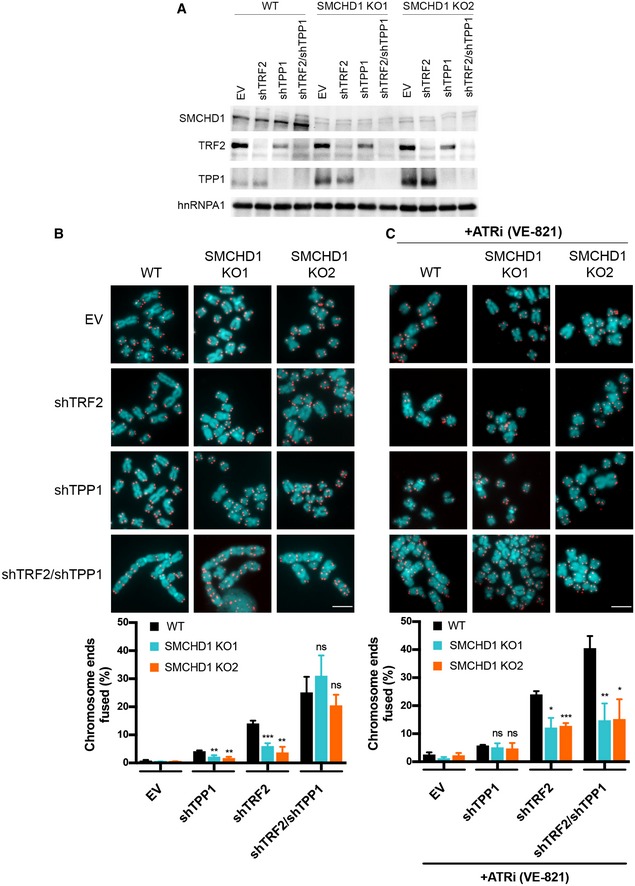
Figure 6. ATR signaling induction by TPP1 removal rescues the telomere fusion defect in SMCHD1 knockout cells.

- Western Blot detection of SMCHD1, TRF2, TPP1 and hnRNPA1 in wild‐type or SMCHD1 knockout HeLa cells transfected with the indicated shRNAs (shTRF2, shTPP1, shTRF2/shTPP1) or EV control.
- Representative metaphase spreads (Scale bar: 5 µm) from HeLa cells transfected with indicated shRNAs or EV control and quantification of telomere fusions. Bars represent average number of fused chromosome ends. SDs were obtained from three independent experiments (> 2,500 telomeres counted/condition/experiment). ***P < 0.001; **P < 0.01 unpaired two‐tailed Student's t‐test.
- Representative metaphase spreads (Scale bar: 5 µm) from HeLa cells transfected with indicated shRNAs or EV control treated for 4 days with ATRi (VE‐821) and quantification of telomere fusions. Bars represent average number of fused chromosome ends. SDs were obtained from three independent experiments (> 2,000 telomeres counted/condition/experiment). ***P < 0.001; **P < 0.01; *P < 0.05 unpaired two‐tailed Student's t‐test.
Source data are available online for this figure.
