
Figure 1.
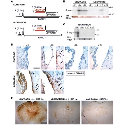
Genome organization, viral spread and blood brain barrier integrity following intracerebral infection with either LCMV Armstrong or rLCMV/INDG. (A) Schema of the LCMV-Armstrong (LMCV-ARM) and rLCMV/INDG genomes. Both viruses consist of two single-stranded negative-strand RNA segments, encoding two viral genes in ambisense orientation each. The long (L) segment encodes for the RNA-dependent RNA polymerase L and for the matrix protein Z, while the short (S) segment carries the GP and NP genes. rLCMV/INDG was created by substituting the LCMV-GP gene for vesicular stomatitis virus-INDG (Pinschewer et al., 2003). (B–E) C57BL/6 mice were infected intracerebrally with rLCMV/INDG or LCMV-ARM as indicated or were left uninfected. (B and C) Viral S-segment (S seg.) RNA in brain was detected at the indicated day (d) after intracerebral infection by northern blot (ethidium bromide staining of 28S rRNA indicates loading control; lanes represent individual animals). (D) Immunohistochemical detection of LCMV-NP confirms reduced spread of rLCMV/INDG as compared to LCMV-Armstrong. Note that LCMV- Armstrong (Days 4 and 6) and also rLCMV/INDG (Day 6) spreads into some subependymal cells (arrows). (E) At Day 6 after infection, animals were given horseradish peroxide (HRP) intravenously. One hour later, they were sacrificed for detection of horseradish peroxidase leakage into the brain parenchyma, indicative of blood brain barrier breakdown. Representative pictures of horseradish peroxidase reactions are shown (n = 3–4 animals per group). Scale bar for D: 100 µm; for E: 500 µm.