
Figure 4.
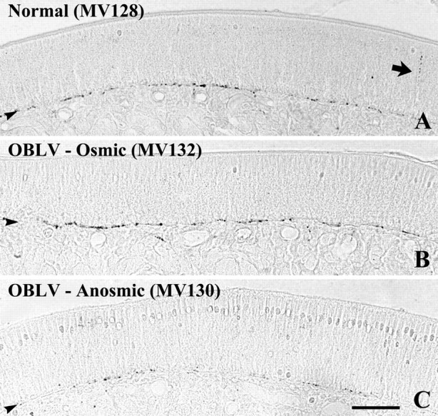
Substance P staining demonstrates differences in the extent of trigeminal fibers across the three behaviorally defined groups. In all three cases, the labeling is concentrated at the basal lamina. (A, B) The density of labeling is roughly equivalent in the normal and OBLV-inoculated, osmic mouse. The arrow in (A) indicates a fiber that ascends through the epithelium toward its apical surface. (C) Substance P-labeled fibers are much reduced in the OBLV-inoculated, anosmic mouse. Scale bar in (C) 50 μm, also applies to (A) and (B).