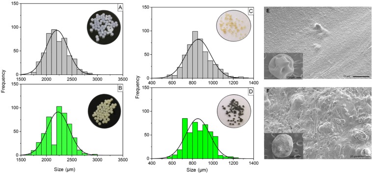
FIGURE 1.
Size distributions of wet and dry calcium alginate microparticles synthesized by ionic gelation, with or without T. harzianum and scanning electron micrographs of the dry calcium alginate microparticles, with and without encapsulated T. harzianum. (A) Wet calcium alginate microparticles; (B) wet calcium alginate/fungus microparticles (1:1 w:w); (C) dry calcium alginate microparticles; (D) dry calcium alginate/fungus microparticles (1:1 w:w). The size distribution was determined using ImageJ software. The wet and dry microparticles presented mean sizes of 2000 and 800 μm, respectively. (E) Calcium alginate microparticles; (F) calcium alginate:fungus microparticles (1:1 w:w). The interiors of the microparticles with and without the fungus showed differences, due to the presence of the fungus (spores) in the second image. The images were acquired at magnifications of 95× (insert) and 250×.

