
FIGURE 1.
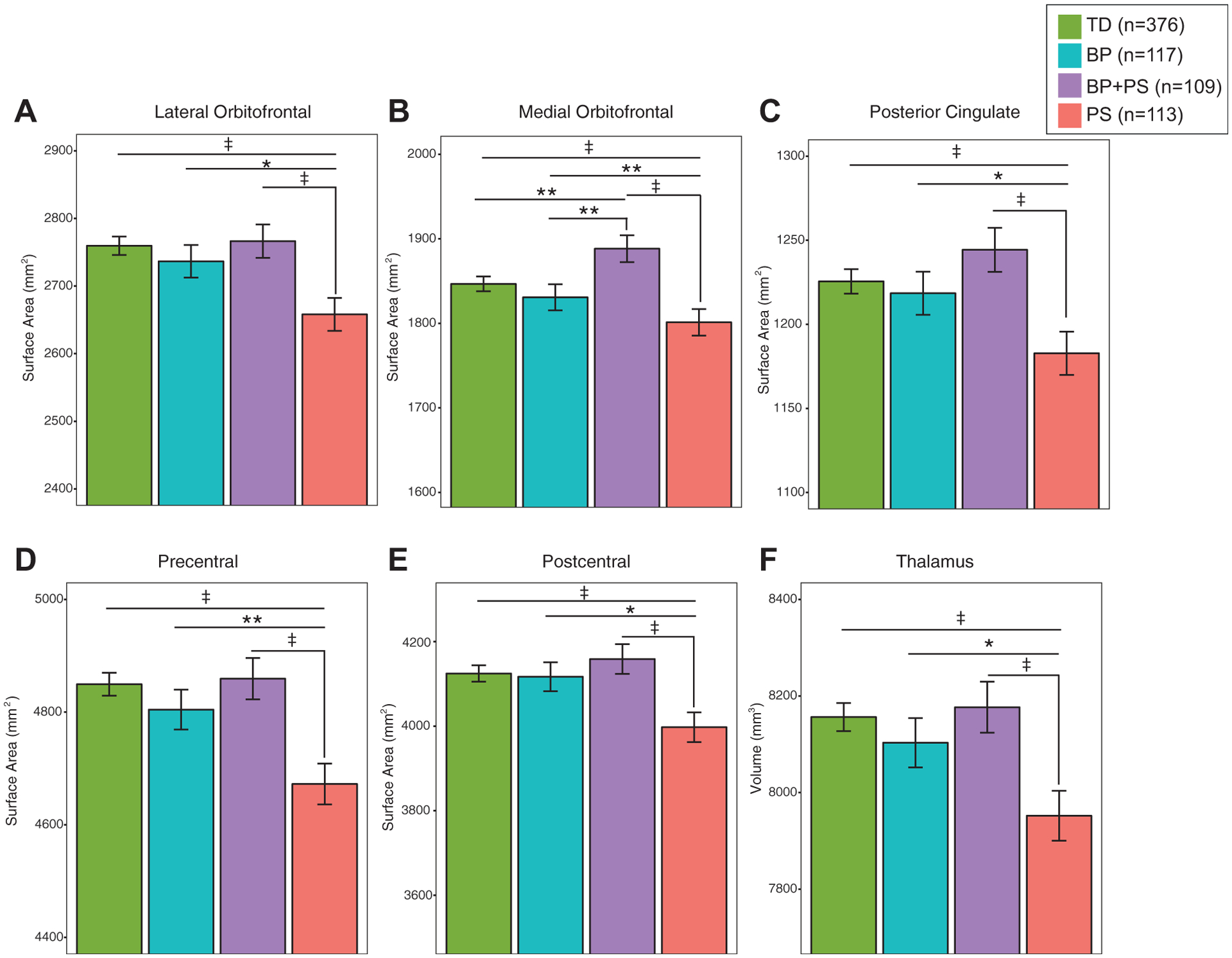
Surface Area Decreases in Psychosis Spectrum (PS) Youth
Note: Psychosis spectrum youth exhibited decreased (A) lateral orbitofrontal, (B) medial orbitofrontal, (C) posterior cingulate, and (D) precentral and (E) postcentral surface areas and (F) thalamic volume compared with typically developing youth (TD), bipolar spectrum youth (BP), and youth with BP and PS.
*p < .05; **p < .01; ‡p < .005.