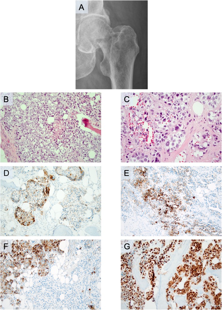
Fig. 2.
Radiological and microscopic findings of the subsequent surgical excision from a pathological hip fracture caused by a metastatic malignant melanoma with neuroendocrine differentiation. All microscopic images are magnified × 200 unless otherwise stated. a. Representative plain radiology scan displaying the pathological hip fracture from which the metastatic melanoma was diagnosed. b. Routine hematoxylin and eosin staining. Note the comedo-like necrosis in the central area. c. Routine hematoxylin and eosin at × 400 magnification, illustrating the nest-forming tumor with nuclear inclusions. d. Focal synaptophysin immunoreactivity (subsets of tumor cells). e. Focal melanoma antigen immunoreactivity (subsets of tumor cells). f. Focal human melanoma black 45 immunoreactivity (subsets of tumor cells). g. Diffuse nuclear SOX10 expression

