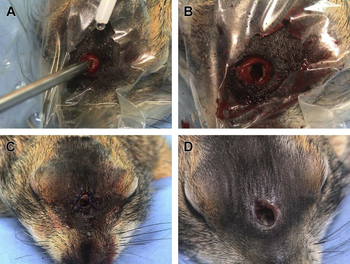
Fig. 17.
Rhinostomy for palliative treatment of pseudo-odontoma in a prairie dog. (A) The anesthetized prairie dog is placed in sternal recumbency. The area over the nasal bones is surgically prepared and draped with a transparent adhesive drape. A circular portion of the skin is removed using a biopsy punch, and the nasal bone is exposed. A 2.5-mm intramedullary pin is used to drill a small rhinotomy access. (B) The rhinotomy site is then enlarged using a small bur. (C) A plastic stent, consisting a of a 1-mL syringe, is placed within the rhinotomy access and sutured to the skin, to keep the rhinostomy site patent. (D) The plastic stent is removed 10 days after the surgical procedure.
(Courtesy of Vittorio Capello, DVM, Milano, Italy; with permission.)

