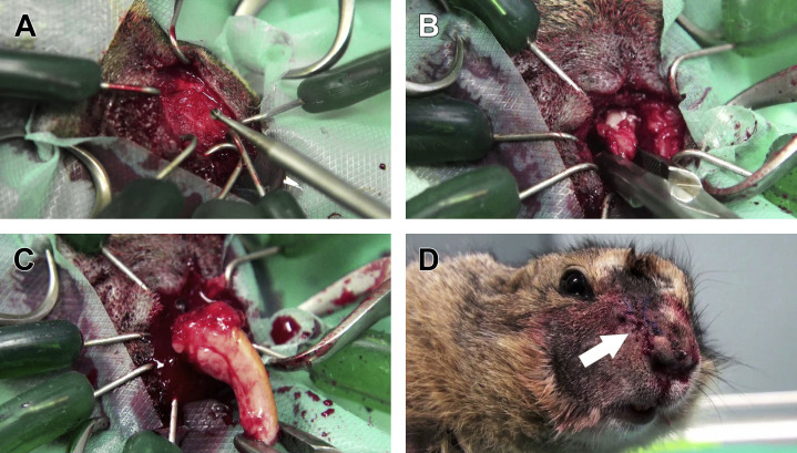
Fig. 19.
Extraction of the right maxillary incisor tooth affected by pseudo-odontoma via the lateral approach to the incisive bone. (A) With the patient anesthetized and placed in lateral recumbency, the lateral aspect of the incisive bone is exposed. Osteotomy is performed with a small 2-mm ball-tipped bur to expose the lateral aspect of the reserve crown of the diseased incisive tooth. (B) Extraction of the rostral portion of the reserve crown. (C) Complete extraction of the diseased tooth, including the deformed apex. (D) Suture of the skin (arrow) and postoperative cosmetic appearance of the surgical access.
(Courtesy of Vittorio Capello, DVM, Milano, Italy; with permission.)

