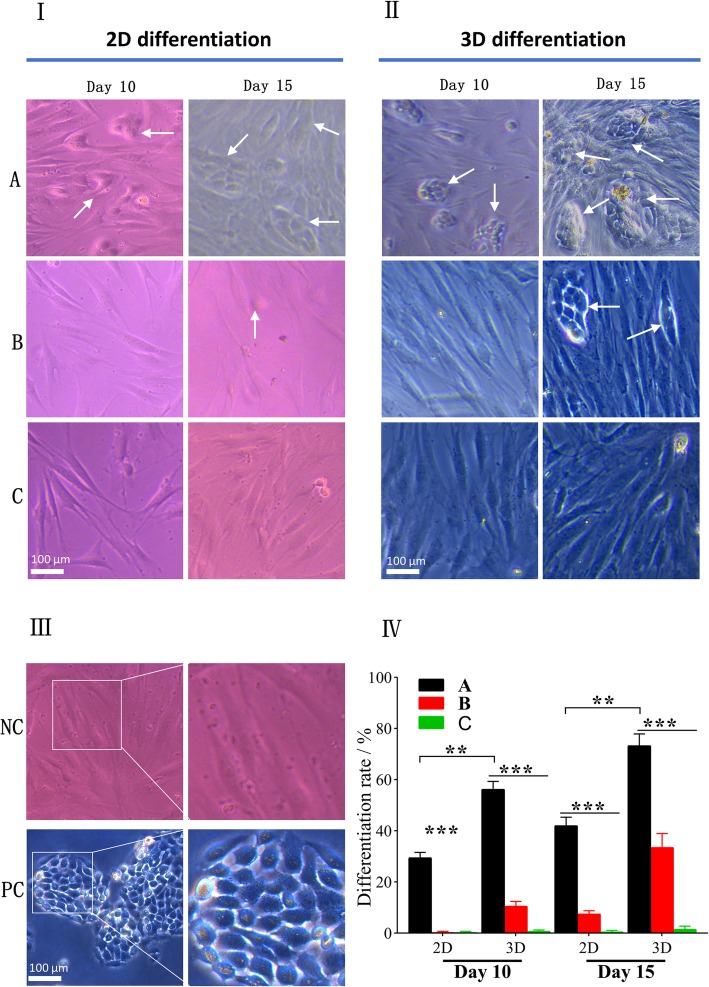
Fig. 4.
Morphology of differentiated hASCs under invert microscope. hASCs with typical cobblestone morphology similar to HaCaT cells were considered differentiated cells (white arrow). I 2D differentiation ((A) hASCs indirectly co-cultured with HaCaT cells at ALI; (B) hASCs indirectly co-cultured with HaCaT cells under ALI; (C) hASCs cultured under ALI without HaCaT’s co-cultivation). II 3D differentiation. III Controls ((NC) undifferentiated hASCs as negative control, (PC) HaCaT cells as positive control). IV Quantification of differentiation rate (%) at different time points (**p < 0.01, ***p < 0.001)

