
Fig. 2.
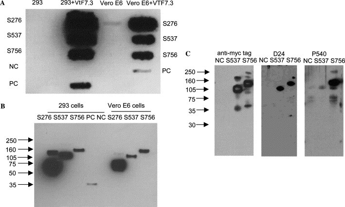
Expression of soluble S fragments. (A) Supernatants of 293 and Vero E6 cells transfected with plasmids encoding S fragments (S276, S537, and S756) in the absence or presence of T7 polymerase expressed by recombinant vaccinia virus (VTF7.3) were transferred to nitrocellulose membranes (dot blots) and detected with anti-c-Myc epitope antibody. PC is the positive control for this antibody provided by the manufacturer and NC is a negative control of cells transfected with empty vector. (B) Supernatants from transfected cells as described above (A) were incubated with Ni–NTA agarose beads, washed, and subjected to Western blotting with the same anti-c-Myc epitope antibody as in A. (C) Detection of S fragments by two rabbit polyclonal antibodies raised against peptides corresponding to sequences starting at residues 24 (D24) and 540 (P540), respectively (right two panels). The left panel shows for comparison Western blot where S537 and S756 were detected by the anti-c-Myc epitope antibody.