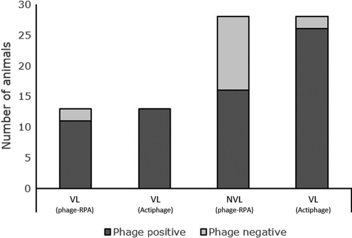
Figure 2.

Comparison of ability of phage‐RPA assay and Actiphage® method to detect M. bovis in blood of cattle. Detection of M. bovis in the blood of cattle stratified by SICCT status and lesion status (visible lesions; VL, or non‐visible lesion, NVL). Phage‐positive samples are in dark grey, and phage‐negative samples are in light grey. The results gained using the Actiphage® method are compared with the previously published results using the phage‐RPA assay (Swift et al., 2016b).
