
Fig. 4.
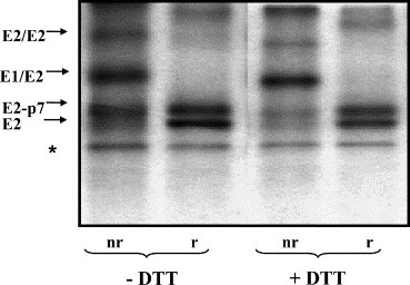
Effect of DTT treatment on the stability of BVDV envelope proteins, after their assembly into dimers. MDBK cells were infected with BVDV at MOI 1. Eighteen hours pi the cells were pulse-labeled with [35S]methionine/cysteine for 15 min and chased for 1 h in the absence of DTT. After this initial chase, 5 mM DTT was added (+) or not (−) to the cells and the incubation continued for 1 h. The cells were lysed and immunoprecipitated with anti-E2 MAb 214. Proteins bound were separated by SDS–10% PAGE under nonreducing (nr) and reducing (r) conditions. The bands marked with an asterisk represent nonspecific contaminating proteins.