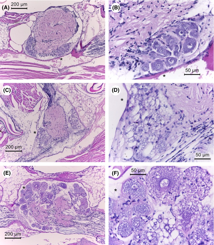
Figure 3.

Photomicrographs of ventral nerve cord ganglia of normal and moribund shrimp from S. khirikhana TH2012T challenge tests. Asterisks (*) indicate the same location in photomicrographs at different magnifications.
A and B. A control shrimp showing normal nerve cord histology at low and high magnification, respectively.
C–F. Moribund shrimp histopathology at progressively higher magnifications showing highly vacuolated cytoplasm of giant nerve cells.
