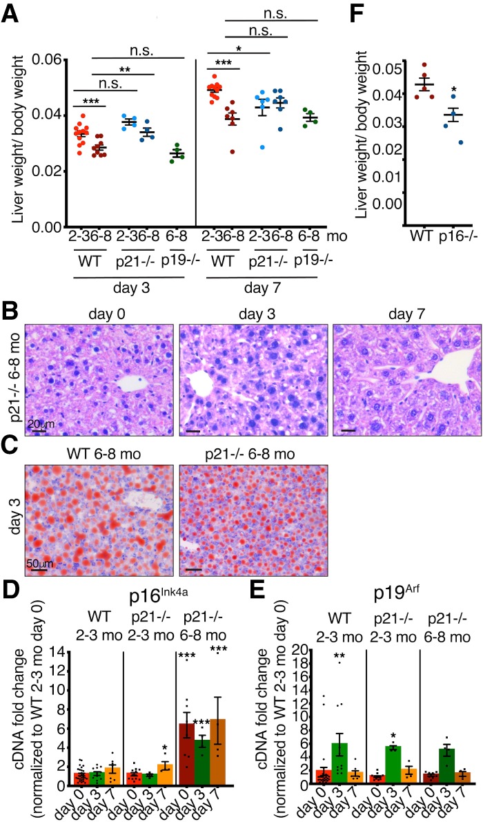
Figure 2.

Adult p21-deficient mice have partially improved regenerative capacity. (A) Liver/body weight ratios of 2- to 3-mo-old and 6- to 8-mo-old WT, p21−/−, and p19−/− mice at 3 and 7 d after PH (n = 4–12). (B) H&E staining of 6- to 8-mo-old p21−/− livers before and at different days after PH. All images are representative of at least four biological replicates. Scale bars, 20 µm. (C) Oil Red O staining of 6- to 8-mo-old WT and p21−/− livers 3 d after PH. All images are representative of at least four biological replicates. Scale bars, 50 µm. (D,E) qPCR analysis for (D) p16Ink4a and (E) p19Arf of 2- to 3-mo-old WT and p21−/− and 6- to 8-mo-old p21−/− livers at different days after PH normalized to 2- to 3-mo-old WT day 0. (n = 4–27). (F) Liver/body weight ratios of 6- to 8-mo-old WT and p16−/− mice 7 d after PH (n = 4–5). Error bars, mean ± SEM, unpaired two-tailed Student's t-test. (*) P ≤ 0.05; (**) P ≤ 0.01; (***) P ≤ 0.001.
