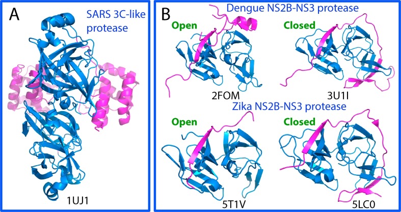
Fig. 1.
Three-dimensional structures of the proteases of SARS-CoV, Dengue and Zika viruses. (A)The dimeric structure of the wild-type 3C-like protease of SARS-CoV. The chymotrypsin folds hosting the catalytic machinery are colored in blue and the extra helical domain in purple. (B) The NS2B-NS3 two component proteases in open and closed forms of Dengue and Zika viruses. The chymotrypsin folds adopted by NS3 protease domain to host the catalytic machinery are colored in blue and the co-factor NS2B in purple.

