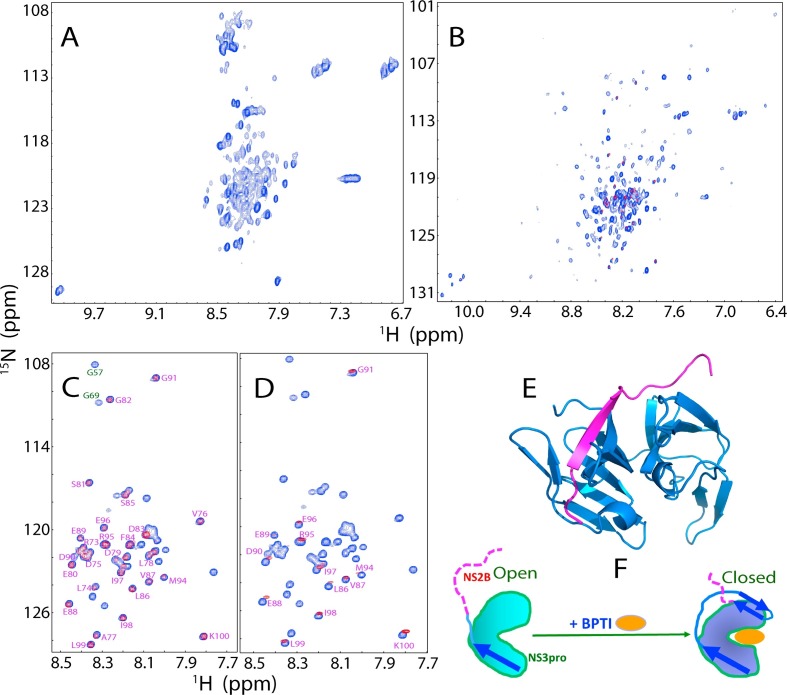
Fig. 9.
The unique properties of the Zika NS2B-NS3 protease. Two-dimensional 1H-15N NMR HSQC spectra of the Zika NS3 protease domain in the isolated state (A), and in complex with the NS2B cofactor (B). (C) Superimposition of HSQC spectra of the 15N-labeled Zika NS2B in the isolated state (blue) and in complex with the unlabeled NS3 protease domain (red). (D) Superimposition of HSQC spectra of the 15N-labeled Zika NS2B in the isolated state (blue), and in complex with unlabeled NS3pro in the presence of unlabelled bovine pancreatic trypsin inhibitor (BPTI) at a molar ratio of 1:2 (red). (E) The crystal structure of the open state of the Zika NS2B-NS3 protease complex (PDB ID of 5T1V). (F) A proposed diagram showing the conformational transformation of NS2B from the open to closed states as triggered by complexing with BPTI. Blue arrows are used for indicating β-strands formed over NS2B, while purple dashed lines are for flexible regions of NS2B.

