
Figure 6.
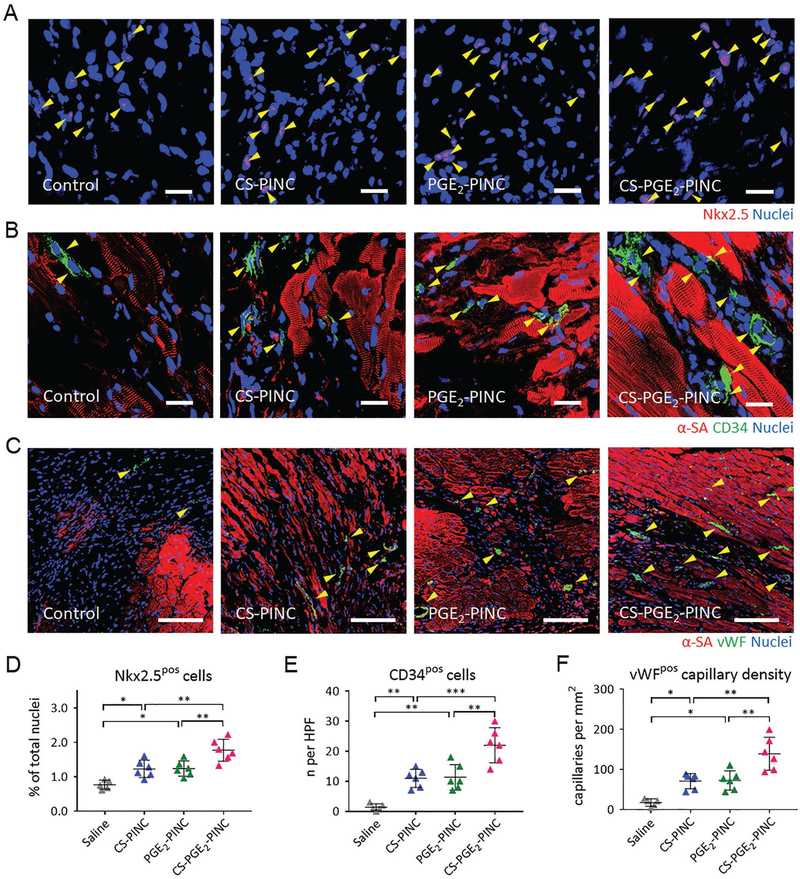
PINC injection promotes endogenous repair in the infarcted heart. A–C) Representative images showing Nkx2.5-positive cells, CD34-positive cells, and vWF-positive capillaries in the infarcted hearts 4 weeks after saline (n = 5), CS-PINC (n = 6), PGE2-PINC (n = 6), or CS-PGE2-PINC (n = 6) treatment. Yellow arrowheads indicate the positively stained cells. D–F) Quantification of the number of D) Nkx2.5-positive cells, E) CD34-positive cells, and F) vWF-positive capillary density in the infarcted hearts 4 weeks after saline (n = 5), CS-PINC (n = 6), PGE2-PINC (n = 6), or CS-PGE2-PINC (n = 6) treatment. Scale bars, A, B) 20 μm; and C) 100 μm. Comparisons among more than two groups were performed using one-way ANOVA followed by post hoc Bonferroni test. * indicates p < 0.05; ** indicates p < 0.01; and *** indicates p < 0.001.