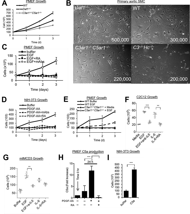
Figure 1. PDGFR and EGFR Signaling.
(A) Primary mouse embryonic fibroblasts (PMEF) (1 ×10⁷) were cultured for 3 d on 10 cm plates in DMEM/F12 and cell numbers quantitated daily. (B) Primary smooth muscle cells (SMC) were isolated from the aortic rings of Daf1−/−, WT, C3−/−Hc−/−, and C3ar1−/−C5ar1−/− mice. Following growth for 2 wks in complete smooth muscle cell media (Lonza) supplemented with 5% FBS, 2 ×10⁶ cells were grown in the same media for 3d after which cells were photographed. (C) Starved PMEFs were incubated for 3 d in DMEM/F12 medium containing 20% FBS, 2 mM L-glutamine, 1% nonessential amino acid, 0.09 mg/ml EC growth supplement, 1% antibiotic/antimycotic, 100 units/ml penicillin, 100 g/ml streptomycin, and 0.09 mg/ml heparin VEGF-A (30 ng/mL) ± anti-C3a/anti-C5a mAbs (5 μg/ml) or C3ar1-A/C5ar1-A (RA; 10 ng/ml ea) and cell numbers were quantified daily. (D) NIH-3T3 cells serum starved in 0.5% serum were incubated over 3 d with PDGF-AA (30 ng/mL) in the absence or presence of anti-C3a/C5a mAbs (5 μg/ml each) or RA (10 ng/ml each and their growth quantified daily. (E) Serum starved cultures of PMEFs from WT and C3ar1−/−C5ar1−/− mice were incubated with media alone or with EGF (30 ng/ml) and cell numbers counted daily. (F) C2C12 myocytes were alternatively cultured for 2 d with EGF (30 ng/ml), EGF+anti-IL-6 (5 μg/ml), IL-6 (10 ng/ml), or IL-6+ RA (10 ng/ml each), after which cell numbers were quantified. (G) mIMCD3 cells were cultured for 3 d with EGF (30ng/ml), EGF+anti-IL-6 (2 μg/ml), IL-6 (10 ng/ml), or IL-6+ RA (10 ng/ml each), after which cell numbers were quantified. (H) PMEFs were incubated for 24 h with media alone or with PDGF-AA (30 ng/ml) in the absence or presence of RA (10 ng/ml each) after which C5a in culture supernatants was assessed by ELISA. (I) NIH-3T3 cells were incubated with media in the absence or presence of added C5a (100 ng/ml) for 3 d after which cell numbers were quantified.

