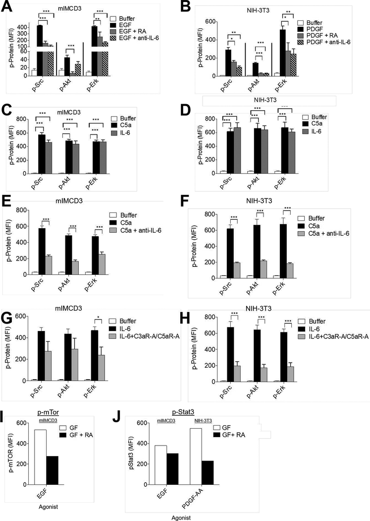
Figure 4. RTK signaling depends on C5ar1/C3ar1 and IL-6R-gp130 co-signaling.
(A) Serum-starved mIMCD3 and (B) serum-starved NIH-3T3 cells were incubated for 5 min with EGF (30 ng/ml) and PDGF-AA (30 ng/ml) respectively in the absence or presence of RA (10 ng/ml each) or anti-IL-6 mAb (10 ug/ml). Following extraction of the cells with the buffer provided by the manufacturer, the extracts were assayed for p-Src, p-Akt, and p-Erk by Luminex (multiplex) assay. (C) Serum-starved mIMCD3 and (D) serum-starved NIH-3T3 cells were incubated for 5 min with media alone, C5a (100 ng/ml) or IL-6 (100 ng/ml). Following extraction of the cells as in panels AB, the extracts were assayed for p-Src, p-Akt, and p-Erk by Luminex (multiplex) assay. (E) Serum-starved mIMCD3, and (F) serum-starved NIH-3T3 cells were incubated for 5 min with C5a (100 ng/ml) or C5a plus anti-IL-6 mAb (10 μg/ml). Following incubation, cells were assayed for p-Src, p-AKT and p-Erk by Luminex (multiplex) assay. (G)Serum-starved mIMCD3 and (H) serum-starved NIH-3T3 cells were incubated for 5 min with IL-6 (10 ng/ml) in the absence or presence of RA (10 ng/ml each. Following incubation for 5 min, cells were assayed for p-Src, p-AKT and p-Erk by Luminex (multiplex) assay. (I) Serum-starved mIMCD3 cells were incubated for 5 min with EGF (30 ng/ml) in the absence or presence of RA (10 ng/ml each). Following incubation for 5 min, cells were assayed for p-mTOR by intracellular FACS staining (system only assays phosphorylated mTOR). (J) Serum-starved mIMCD3 or serum-starved NIH-3T3 cells were incubated for 8 h with EGF (30 ng/ml) or PDGF-AA (30 ng/ml) respectively in the absence of presence of RA (10 ng/ml each), after which, phospho-STAT3 was assayed by flow cytometry (system only assays phosphorylated STAT3).

