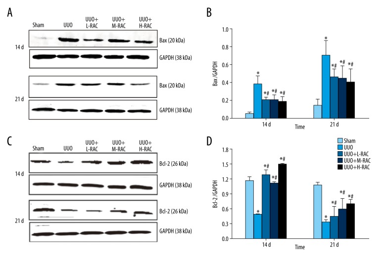
Figure 7.
Effect of RAC on the expression of Bax and Bcl-2 proteins in rat renal tissues detected by immunoblotting. The total protein was extracted from the renal tissue sample from each group. Equal amounts of proteins were separated by western-blotting analyses. The expression of Bax (A) and Bcl-2 (C) protein were determined by using rabbit-anti-rat Bax and rabbit-anti-rat Bcl-2 antibodies, respectively. The result of western blotting was then quantitated by calculating and analyzing the gray image values. The relative expression of (B) Bax and (D) Bcl-2 in renal tissues from Sham, UUO, UUO+L-RAC, UUO+M-RAC, and UUO+H-RAC groups of rats are shown. Data are presented as means±SD (n=10 in each group). * P<0.05 compared to the Sham group. # P<0.05 compared to the UUO group. All results were assessed by 3 independent experiments. RAC – rhubarb and astragalus capsule; TGF – transforming growth factor; UUO – unilateral ureteral obstruction; SD – standard deviation.

