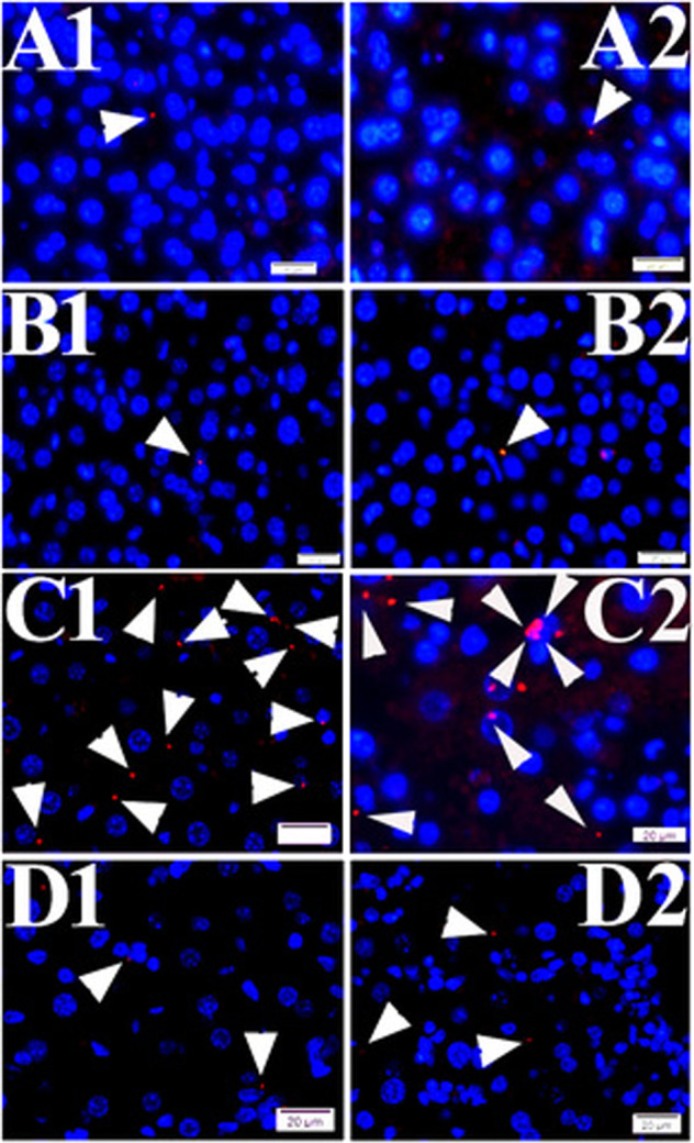
Fig. 6. PLA signals of TVSPs in liver tissues from WT and EPAC1 KO mice.

PLA signals (red) of TVSPs (arrowheads) in liver tissues from wild-type (WT) mock (a1, a2), EPAC1-KO mock (b1, b2), WT-R. australis (c1, c2), and EPAC1-KO R. australis-infected mice (d1, d2). Nuclei were counter-stained with DAPI (blue). Scale bars indicate 20 µm.
