
Fig. 7.
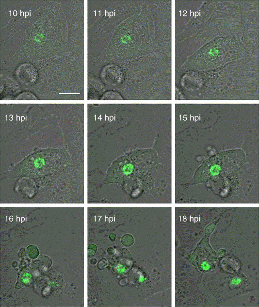
Live-cell analysis of the last stages of B54GFP-2 infection. Series of EGFP and transmitted light simultaneous acquisitions of the same B54GFP-2 infected Vero cell by confocal microscopy from 10 hpi (when viral factory is already constituted) and every 5 min (see additional file 3 to complete animation sequence). Rounding of the infected cell is followed by membrane blebbing and subsequent formation of p54-EGFP (green) containing vesicles. At 18 hpi, detachment and final cell destruction is observed. Scale bar, 20 μm. (For interpretation of the references to colour in this figure legend, the reader is referred to the web version of this article.)