
Fig. 1.
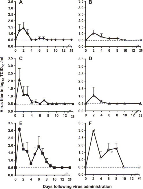
SARS-CoV replication in the respiratory tract of monkeys. Mean titers of virus (expressed as log10 TCID50/ml of sample; y axis) detected on indicated days (x axis) in the upper respiratory tract (left panels, A, C, and E, closed symbols) and lower respiratory tract (right panels, B, D, and F, open symbols) of rhesus (panels A and B, ◆, ◇), cynomolgus (panels C and D, ▴, ▵), and African Green (panels E and F, ■, □) monkeys following intranasal and intratracheal administration of 106 TCID50 of SARS-CoV. Error bars associated with each data point indicate standard error, and the dotted line indicates the lower limit of detection of virus (100.5 TCID50/ml).