
Fig. 3.
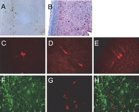
Neurons are the primary target of HCoV-OC43NV in vivo. Mice were inoculated intranasally with 30 LD50 of HCoV-OC43NV and brains and spinal cords were harvested 7 or 9 days p.i. Tissue samples were prepared for in situ hybridization, antigen staining or Luxol Fast Blue (LFB) staining and slides were examined with standard light, fluorescence or confocal microscopy as described in Materials and methods. (A–B) HCoV-OC43 antigen is localized in the gray matter of spinal cords. Panel A depicts immunohistochemical detection of HCoV-OC43 using the anti-OC43 S hybridoma O.4.3. Panel B depicts a serial section stained with LFB to demarcate the white matter. (C–E) Morphology of HCoV-OC43NV-infected cells is consistent with that of neurons. Images are representative coronal sections of brain stems from mice harvested 7–9 days p.i. Sections were stained with O.4.3 followed by Cy3-labeled goat anti-mouse. (F–H) Combination in situ hybridization for HCoV-OC43 nucleocapsid RNA (Cy3-labeled antisense probe, red) and immunohistochemistry for the astrocyte marker GFAP (FITC-labeled anti-GFAP, green). Panel H is a merged image of panels F and G. Original images are 20× magnification for panels A and B, or 40× magnification for panels C–H.