
Fig. 4.
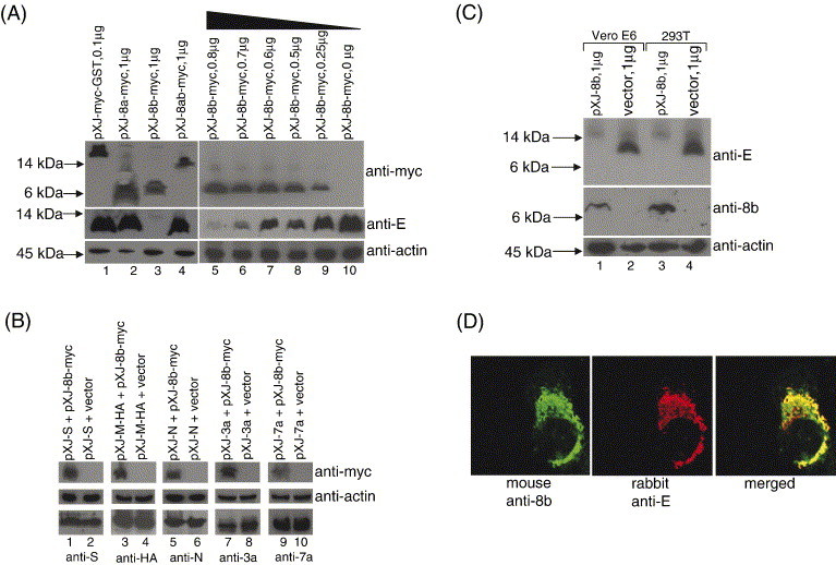
Effects of 8b on the expression of the small structural protein, E. (A) 293T cells were co-transfected with 2 μg of pXJ-E and 0.1 μg of pXJ-myc-GST (lane 1), 1 μg of pXJ-8a-myc (lane 2), pXJ-8b-myc (lane 3), pXJ-8ab-myc (lane 4), or decreasing amount of pXJ-8b-myc (lanes 5–10). Total cell lysates were subjected to Western blot analysis to determine the expression of E (middle panel) and myc-tagged proteins (top panel). Equal amounts of cells were used in each lane as verified by the level of endogenous actin (bottom panel). (B) 293T cells were co-transfected with 1 μg of pXJ-S and 1 μg of either pXJ-8b-myc or empty vector (lanes 1 and 2). Total cell lysates were subjected to Western blot analysis to determine the expression of 8b-myc (top panel) and S (lower panel). Equal amounts of cells were used in each lane as verified by the level of endogenous actin (middle panel). Similar experiments were performed with 1 μg of pXJ-M-HA (lanes 3 and 4), 0.25 μg of pXJ-N (lanes 5 and 6), 0.4 μg of pXJ-3a (lanes 7 and 8) or 0.4 μg of pXJ-7a (lanes 9 and 10). (C) Vero E6 or 293T cells were co-transfected with 2 μg of pXJ-E and 1 μg of pXJ-8b (lanes 1 and 3) or 1 μg of empty vector (lanes 2 and 4). Total cell lysates were subjected to Western blot analysis to determine the expression of E (top panel) and 8b (middle panel). Equal amounts of cells were used in each lane as verified by the level of endogenous actin (bottom panel). (D) Indirect immunofluorescence experiments were performed to determine the cellular localization of 8b and E in Vero E6 cells co-transfected with pXJ-8b and pXJ-E. The expression of 8b is represented by FITC staining (left panel), whereas the expression of E is represented by rhodamine staining (middle panel). The merged images showed that the 8b and E partially colocalized in co-transfected cells (right panel).