
Fig. 5.
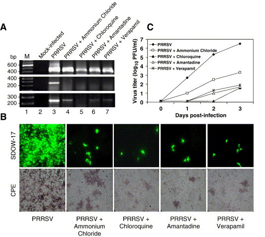
Inhibition of PRRSV replication by ion channel blockers. (A) Strand-specific RT-PCR for N gene. The drug concentrations were used according to the previously report (Kreutz and Ackermann, 1996). Verapamil concentration was determined by limiting dilution for cell cytotocixity in Marc-145 cells. Marc-145 cells were treated with ammonium chloride (5 mM), chloroquine (20 μM), amantadine (1 mM) or verapamil (50 μM) for 30 min prior to PRRSV infection and the virus-infected cells were incubated for 3 days in the presence of the drug. Total RNA was extracted from cells at 2 days (upper and middle panels) or 3 days (lower panel) post-infection, and strand-specific RT-PCR was conducted to detect positive-sense (upper panel) or negative-sense (middle and lower panels) genome. Lane 1, molecular weight marker; lane 2, mock-inoculated cells; lane 3, untreated; lanes 4 to 7, treated with the indicated drug. (B) PRRSV replication in the presence of the drug. Virus-infected, drug-treated Marc-145 cells were stained for immunofluorescence (upper panels) with N-specific MAb SDOW-17 at 2 days post-infection (magnification 20×). PRRSV-specific CPE was monitored and photographed at 3 days post-infection (lower panels). (C) Growth kinetics of PRRSV in the presence of drugs. Cells infected with PRRSV were incubated in the presence of the drug, and culture supernatants were harvested at the indicated times for plaque assays in duplicate.