
Fig. 3.
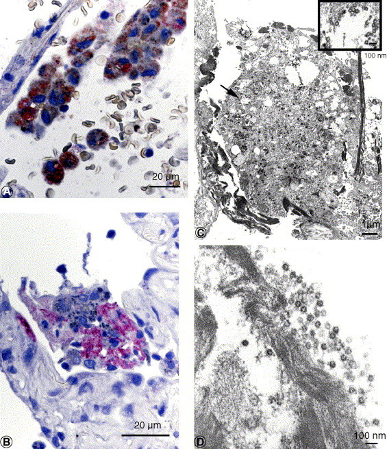
A, SARS-CoV and CD68 antigens in alveolar macrophage. Red stain, SARS-CoV; brown stain, CD68 (double-stain IHC with immunoalkaline phosphatase and peroxidase polymer). B, SARS-CoV antigens in intraalveolar necrotic debris. C and D, Extracellular virions were often associated with fibrin within the alveolar space. C inset, Higher magnification of area at arrow. Bar size indicated in micrometers or nanometers.