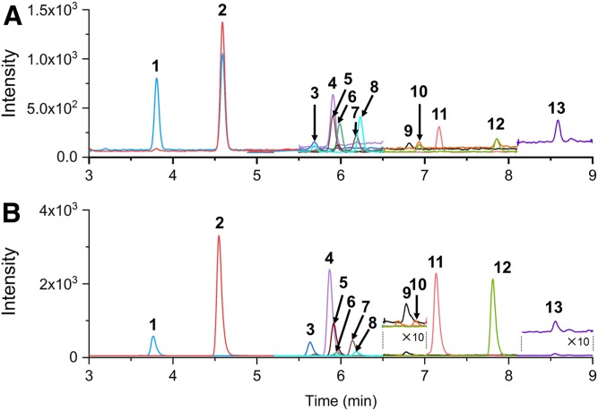
Fig. 5.
Chromatograms for the separation of steroids with 100 pg being injected: without LiCl (H method) (A); with LiCl (Li method) (B) (see the Materials and Methods). 1, 16-OH-E1; 2, 7-OH-DHEA; 3, TH-COL; 4, 7-OH-P5; 5, TH-COR; 6, 11-OH-An; 7, THB; 8: APD; 9, DHEA; 10, 17-OH-P5; 11, TH-DOC; 12, THS; 13, P5. The MRM transitions used in this analysis were shown in supplemental Tables S1 and S2. The peak traces depicted in colors correspond to those obtained by the transitions indicated in bold, which were used for the quantification.

