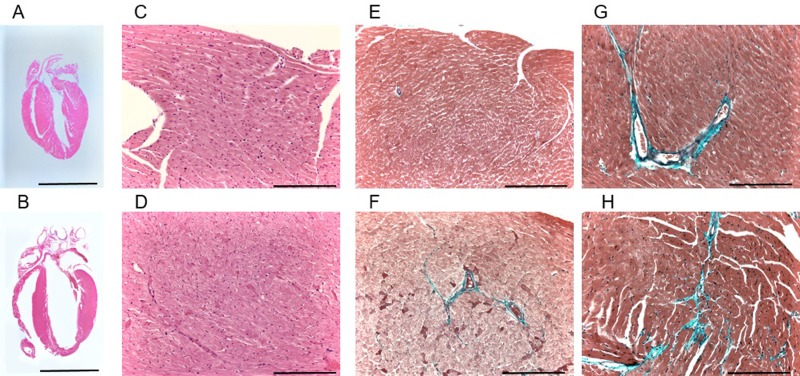
Fig 1. Histological findings in wild-type (upper panels) and TBX5R264K/R264K mice (lower panels).
A and B. Longitudinal sections through the atria and ventricles with hematoxylin-eosin staining. Scale bars: 2 mm. C and D. Hematoxylin-eosin staining of high magnification sections of left ventricle myocardium. Scale bars: 100 μm.E and F. Elastica-Masson staining of sections from the left ventricular apex to free wall of hearts from young adult mouse. Slight fibrosis (blue signals) is seen around the vessels in young adult TBX5R264K/R264K mouse. Scale bars: 100 μm. G and H. Elastica-Masson staining of sections from the left ventricular apex to free wall of hearts from mature to middle age mice. Mild fibrosis (blue signals) from the endocardium to gaps between cardiomyocytes, and increased perivascular fibrosis with extension to the interstitium are shown in TBX5R264K/R264K compared with wild-type. Scale bars: 100 μm.

