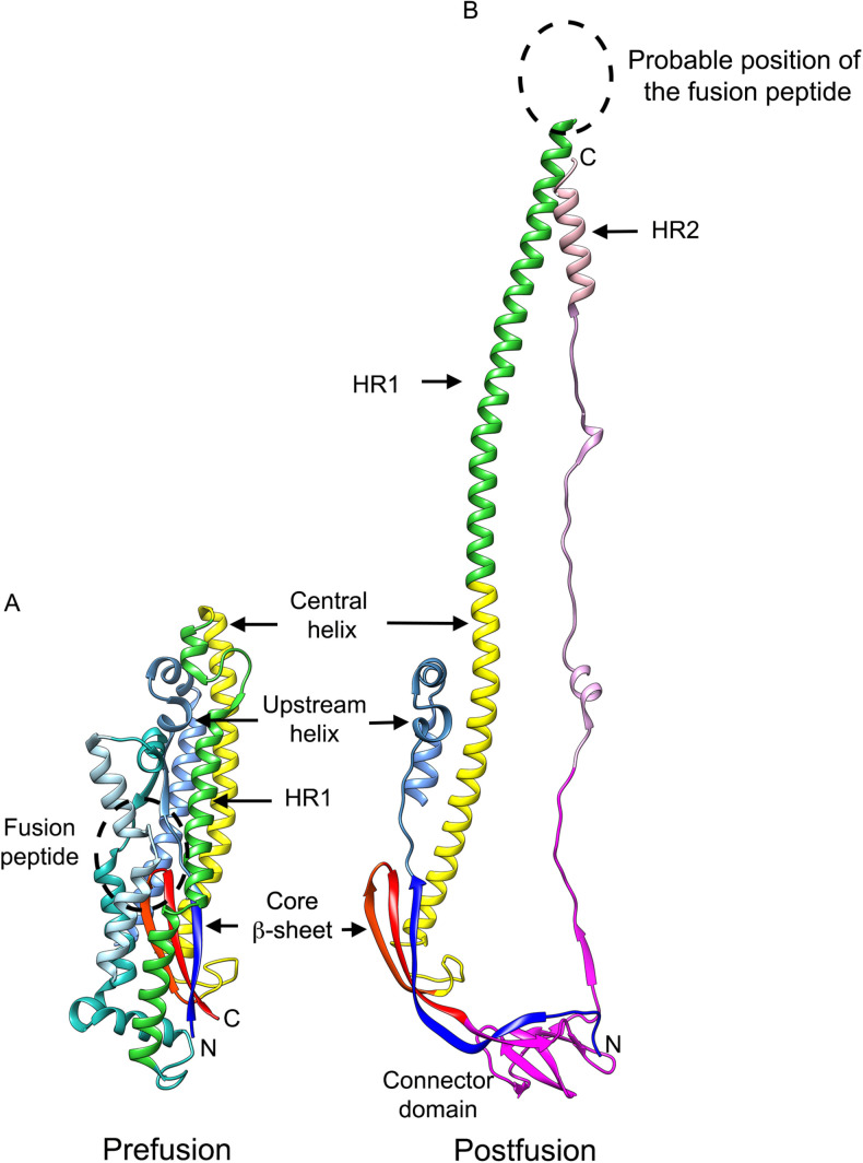
Fig. 3.
CoV S conformational changes driving the fusion reaction. (A), Ribbon diagram of the MHV S2 subunit in the prefusion conformation, PDB: 3JCL. (B), Ribbon diagram of the MHV S2 subunit in the postfusion conformation, PDB: 6B3O. The prefusion to postfusion transition involves a “jack-knife” refolding of the HR1 helices and intervening regions into a single continuous helix appended to the central helix. The connector domain and HR2 in the prefusion structure and the fusion peptide in the postfusion structure of MHV were not resolved and are therefore not shown.

