
Fig. 4.
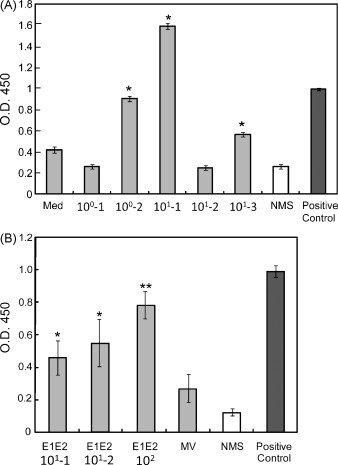
Detection of human MV-specific antibodies in the serum of rMV- or rMV-E1E2-infected mice. (A) Serum (1:100) from MV-infected mice (100–101 pfu) was analysed by ELISA using an MV-infected B95a cell lysate as the target. An anti-MV-NP antibody was used as a positive control and NMS indicates normal mouse serum. The asterisk (*) indicates a significant reaction (p < 0.01) compared to the medium alone control. (B) Serum (1:100) from rMV-E1E2-infected mice (101–102 pfu) was analysed by ELISA. An anti-MV-NP antibody was used as a positive control and NMS indicates normal mouse serum. The double asterisk (**) indicates a highly significant reaction (p < 0.001) compared to NMS and a single asterisk (*) indicates a significant reaction (p < 0.05) compared to NMS.