
Fig. 4.
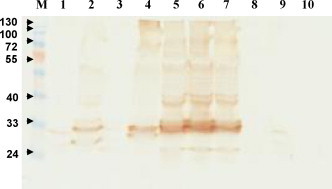
Expression of scFv antibodies analyzed by Western blotting. Identical amounts of total cellular lysates from 30 clones were loaded onto SDS-PAGE and transferred to nitrocellulose papers. The presence of scFv antibodies was detected by goat anti-chicken light chain antibodies at 1:3000 dilution, followed by HRP-conjugated donkey anti-goat IgG. The predicted molecular weight of scFv fragment is approximately 35 kDa. The blot is a representative result of scFv expression in 10 selected clones.