
Fig. 8.
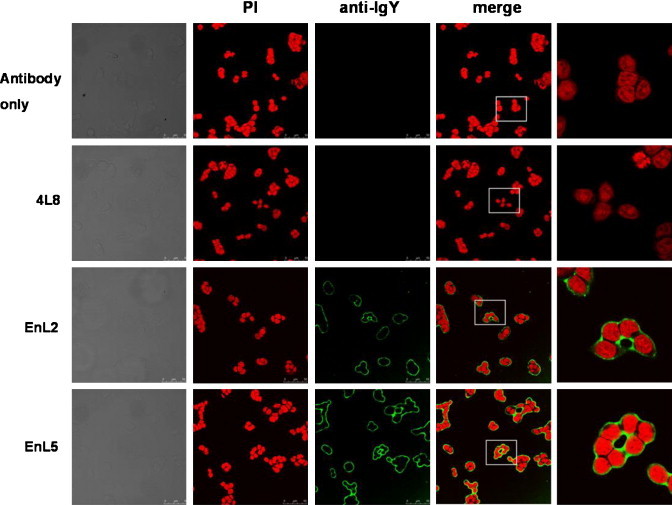
Immunofluorescent staining of α-enolase protein in PE089 cells. Cells were fixed and their α-enolase expression was detected using purified EnL2 and EnL5 scFv antibodies, followed by mouse anti-HA and Cy-2-conjugated goat anti-mouse antibodies. The nucleus (red) was visualized by PI staining. Both EnL2 and EnL5 scFv antibodies clearly stained nuclear membrane (green) in PE089 cells. An anti-SARS-CoV scFv antibody, 4L8, did not show any reactivity with nuclear membrane. (For interpretation of the references to color in this figure legend, the reader is referred to the web version of the article.)