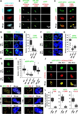
Fig. 3. ENL promotes the multivalent phase separation of SEC.

(A) Live cell imaging of HeLa cells coexpressing eCFP-CCNT1 and CDK9-mRFP. (B) Fluorescence microscopy images showing that the purified mCherry-CDK9 proteins can form heterotypic droplets together with AFF4-IDR-eGFP or ENL-IDR-eGFP. The purified eGFP protein was used as a negative control. Purified proteins (10 μM) were used, and the droplet formation buffer contains 10% PEG-8000 and 50 mM NaCl. (C) Live cell imaging of HeLa cells expressing CDK9-mRFP together with eGFP-AFF4 or eGFP-ENL. (D) IF imaging of CDK9 in wild-type and ENL knockout HCT 116 cells. DNA was counterstained using DAPI. (E) Box plot showing that the number of CDK9 puncta per nucleus is significantly decreased after ENL knockout. Each n > 30 nuclei; error bars represent the distribution between the 90th and 10th percentiles. Results are representative of three biological replicates. (F) IF imaging of AFF4 in wild-type and ENL knockout HCT 116 cells. DNA was counterstained using DAPI. (G) Box plot showing that the number of AFF4 puncta per nucleus is significantly decreased after ENL knockout. Each n > 30 nuclei; error bars represent the distribution between the 90th and 10th percentiles. Results are representative of three biological replicates. (H) Live cell imaging of HCT 116 wild-type and ENL knockout cells expressing eGFP-AFF4. (I) Box plot showing that the number of eGFP-AFF4 droplets per nucleus is significantly decreased after ENL knockout, which can be rescued by overexpression of mCherry-ENL. Each n > 20; error bars represent the distribution between the 90th and 10th percentiles. Results are representative of three biological replicates. (J) Live cell imaging of HCT 116 wild-type and ENL knockout cells coexpressing eGFP-AFF4 and mCherry-CCNT1. (K) IF imaging showing costaining of AFF4 with ENL, CDK9, or ELL2 in HeLa cells transfected with MLL-ENL (left) or ENL (right). DNA was counterstained using DAPI. (L) Box plot showing that the number of AFF4/ENL (left), AFF4/CDK9 (middle), and AFF4/ELL2 (right) colocalized puncta per nucleus is significantly increased after transfection with MLL-ENL. Each n > 20; error bars represent the distribution between the 90th and 10th percentiles. Results are representative of three biological replicates.
