
Fig. 1.
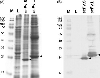
Total cellular lysates seen using Coomassie blue dye (A), while single-chain variable fragment (scFv) antibodies were shown by the western blotting technique (B). The recombinant scFv.S (with short linker) and scFv.L (with long linker) antibodies were identified as 24 and 28 kDa bands, respectively. Total cellular lysates from XL-1 bacteria were loaded into lane L. Lane M denotes prestained protein markers.