
Fig. 1.
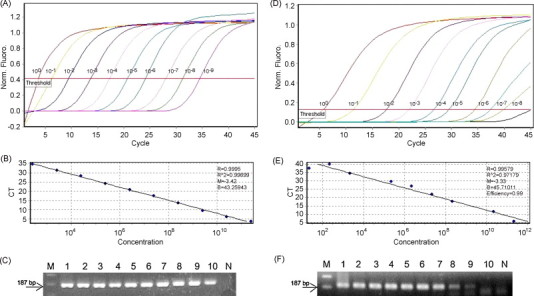
Standards for the SYBR Green real-time RT-PCR assay for the quantitation of BToV and PToV cRNA. (A) Amplification of 100, 10−1, 10−2, 10−3, 10−4, 10−5, 10−6, 10−7, 10−8 and 10−9 dilutions of the cRNA standard used in parallel with each SYBR Green-based real-time RT-PCR assay. (B) Standard curves of the real-time RT-PCR based on serial dilutions of BToV cRNA standards. In the standard curve of these dilutions, each dot represents the result of duplicate amplification of each dilution. The coefficient of determination (R2) and the slope(s) of the regression curve are indicated. (C) SYBR Green real-time RT-PCR products using serially diluted in vitro transcripts. M, molecular marker; lanes 1–10: 2.54 × 10¹¹, 2.54 × 10¹⁰, 2.54 × 10⁹, 2.54 × 10⁸, 2.54 × 10⁷, 2.54 × 10⁶, 2.54 × 10⁵, 2.54 × 10⁴, 2.54 × 10³ and 2.54 × 10² viral copies/reaction; N, negative control. (D) Amplification of 100, 10−1, 10−2, 10−3, 10−4, 10−5, 10−6, 10−7, 10−8 and 10−9 dilutions of cRNA standard used in parallel with each SYBR Green-based real-time RT-PCR assay. (E) Standard curves of the real-time RT-PCR based on serial dilutions of PToV cRNA standards. In the standard curve of these dilutions each dot represents the result of duplicate amplification of each dilution. The coefficient of determination (R2) and the slope (s) of the regression curve are indicated. (F) SYBR Green real-time RT-PCR products using serially diluted in vitro transcripts. M, molecular marker; lanes 1–10: 2.17 × 10¹¹, 2.17 × 10¹⁰, 2.17 × 10⁹, 2.17 × 10⁸, 2.17 × 10⁷, 2.17 × 10⁶, 2.17 × 10⁵, 2.17 × 10⁴ and 2.17 × 10³ viral copies/reaction; N, negative control.