
Fig. 2.
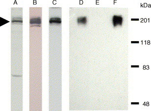
Western blot analysis to detect S-EGFP from CHO-SG cell lysate and samples prepared by using WGA affinity chromatography. Protein samples were resolved by 8% SDS PAGE and then transferred to a nitrocellulose membrane. Lanes A–C are the identification of S-EGFP in CHO-SG cell lysate. The same membrane was probed with three antibodies separately. Lane A, mouse monoclonal anti-GFP antibody; lane B, rabbit anti-S serum; and lane C, horse anti-S serum. S-EGFP is indicated by the arrowhead. Lanes D–F are the results of WGA chromatography. Lane D, CHO-SG cell lysate; lane E, the flow-through from the WGA column; and lane F, sample eluted from the WGA column. Rabbit anti-S antibody was used for lanes D–F.