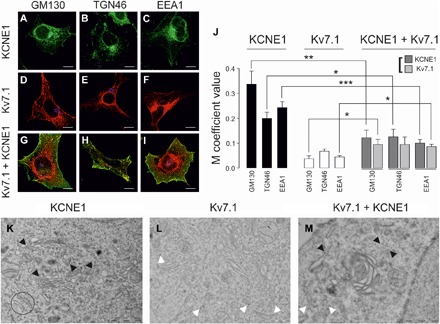
Fig. 4. Kv7.1 and KCNE1 colocalization with post-ER compartments of the secretory pathway.

(A to C) KCNE1 colocalization with post-ER subcellular compartment markers. (A) Cis-Golgi network (GM130), (B) TGN (TGN46), and (C) early endosomes (EEA1). Green, KCNE1-YFP; blue, subcellular compartments; cyan, colocalization between green and blue. (D to F) Kv7.1 colocalization with GM130 (D), TGN46 (E), and EEA1 (F). Red, Kv7.1-CFP; blue, subcellular compartments; magenta, colocalization between red and blue. (G to I) Kv7.1 + KCNE1 colocalization with GM130 (G), TGN46 (H), and EEA1 (I). KCNE1-YFP localization is shown in green, Kv7.1-CFP is shown in red, and the corresponding subcellular compartments are shown in blue. White shows triple colocalization between green, red, and blue. Yellow shows partial green and red colocalization. Scale bars, 10 μm. (J) The values represent the means ± SEM of the M overlap coefficient between KCNE1 (black), Kv7.1 (white), or Kv7.1 + KCNE1 (light and dark gray) and the corresponding subcellular compartment. *P < 0.05, **P < 0.01, and ***P < 0.001 (n = 10 to 34, Student’s t test). (K to M) Electron micrographs of KCNE1 (K), Kv7.1 (L), and Kv7.1 + KCNE1 (M) in HEK-293 cells. (K) KCNE1 staining of Golgi cisternae as indicated by the black arrowheads. Representative ER structures are circled. (L) Kv7.1 was located in ER-like structures, indicated by the white arrowheads, and was absent from the vicinity of the Golgi. (M) KCNE1 (indicated by the black arrowheads) disappeared from the Golgi cisternae and was located in ER-like structures in the presence of Kv7.1 (indicated by the white arrowheads). Kv7.1, 12-nm gold particles; KCNE1, 18-nm gold particles. Scale bars, 1 μm.
